College of Medicine – Tucson researchers used stem cell technology to improve research models for Parkinson’s disease.

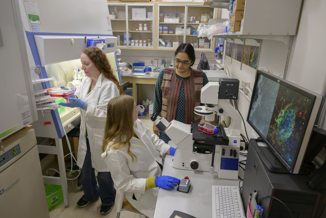
Lalitha Madhavan, MD, PhD, and her research team used induced pluripotent stem cell technology to reprogram adult skin cells into brain cells to study Parkinson’s disease. (University of Arizona Health Sciences)
A recent study published in Progress in Neurobiology and led by researchers at the University of Arizona College of Medicine – Tucson has developed an improved method to study Parkinson’s disease in the lab. Along the way, researchers also uncovered clues that may help scientists figure out how to detect Parkinson’s earlier and point the way toward better treatments.
Around a million Americans are living with Parkinson’s disease, a neurological disorder that causes difficulty in movement, balance and cognition. Symptoms worsen until tasks like walking, talking and swallowing present enormous challenges. While there is no cure, there are treatments that control symptoms — but their effectiveness wanes over time and they are associated with unwanted side effects.
“It’s a slow-developing disorder. We only diagnose the disease at a late stage, when 60-70% of dopamine neurons are dysfunctional or have died off,” said Lalitha Madhavan, MD, PhD, associate professor of neurology at the College of Medicine – Tucson, part of UArizona Health Sciences. “We have treatments, but at that point you’re trying to throw a small glass of water on a raging fire. Being able to diagnose the condition at the earliest stages would be a big step.”
Madhavan’s team used cells from Parkinson’s patients to create a human-derived laboratory model to study the disease. Using induced pluripotent stem cell technology — a powerful technique that transforms adult cells into embryo-like cells that can then mature into any cell type — the lab reprogrammed adult skin cells called fibroblasts into brain cells.
Using the reprogrammed neurons, Madhavan Lab researchers discovered several changes in the cells from Parkinson’s subjects that differentiated them from cells of healthy individuals. Madhavan hopes this finding can form the basis for better cell-culture systems for studying Parkinson’s disease in the lab, potentially leading to improved diagnostics and treatments.
The experiments also showed that skin cells may act as a window into the brain. Skin cells don’t cause neurological symptoms, but some of the same changes that damage brain cells might also affect skin cells, producing similar molecular “signatures.”
“We wanted to make neurons from skin biopsies using this fantastic technology; however, we noted along the way that the fibroblasts themselves seemed to have signatures that differentiated individuals with Parkinson’s. We started to dig deeper into that,” Madhavan said. “It’s exciting that we’ve shown that connection, and that it tells us skin cells could perhaps be used to diagnose the disease early.”
The team hopes that, in the future, doctors will be able to catch Parkinson’s disease earlier by examining skin cells for signs that the disease is brewing.
“This could be a system in which we could very carefully diagnose people at early stages,” Madhavan said, adding that her team received a patent on a method for examining skin cells for molecular signs that correlate to Parkinson’s disease.
They are now investigating how skin cells change over time to learn more about how the disease progresses and how to identify it early. Tech Launch Arizona, the University of Arizona’s technology commercialization office, is helping protect the innovation and developing strategies to take it from the laboratory to the marketplace where it can impact the lives patients and their doctors.
Madhavan says that if we could catch Parkinson’s disease earlier, doctors could prescribe currently available treatments that can slow disease progression. Simultaneously, scientists could work to develop next-generation Parkinson’s drugs that target the disease in its early stages.
Because a patient’s skin cells are easy to access — especially compared to brain cells — Madhavan also hopes the system could be used for a precision-medicine approach, matching patients with optimized treatments based on a skin biopsy and lab test showing which drug might work best based on their unique genetic profile.
“We’ve been putting Parkinson’s into one big bucket when actually different people express it differently,” she said. “This system would allow us to carefully classify Parkinson’s and assess treatments more effectively based on such a classification.”
The lead authors on the study were Mandi Corenblum, MS, senior research specialist, and Aiden McRobbie-Johnson, physiological sciences graduate student. Co-authors include Kelsey Bernard and Timothy Maley, graduate students in neuroscience and physiological sciences; Emma Carruth, undergraduate student in physiology; Moulun Luo, PhD, associate research professor of medicine; Lawrence Mandarino, PhD, professor of medicine; Maria Sans-Fuentes, PhD, BIO5 Institute statistician; Dean Billheimer, PhD, professor in the UArizona Mel and Enid Zuckerman College of Public Health and director of statistical consulting at the BIO5 Institute; and Erika Eggers, PhD, professor of physiology and member of the BIO5 Institute.
The study was supported mainly by a Michael J Fox Foundation grant (MJFF 18366) and in part by grants from the National Eye Institute, a division of the National Institutes of Health, under award nos. R01EY026027 and NSF1552184.
Contacts
Anna Christensen, College of Medicine – Tucson
520-626-9964, achristensen@arizona.edu
David Bruzzese, College of Medicine – Tucson
520-626-9722, dbruzzese@arizona.edu
